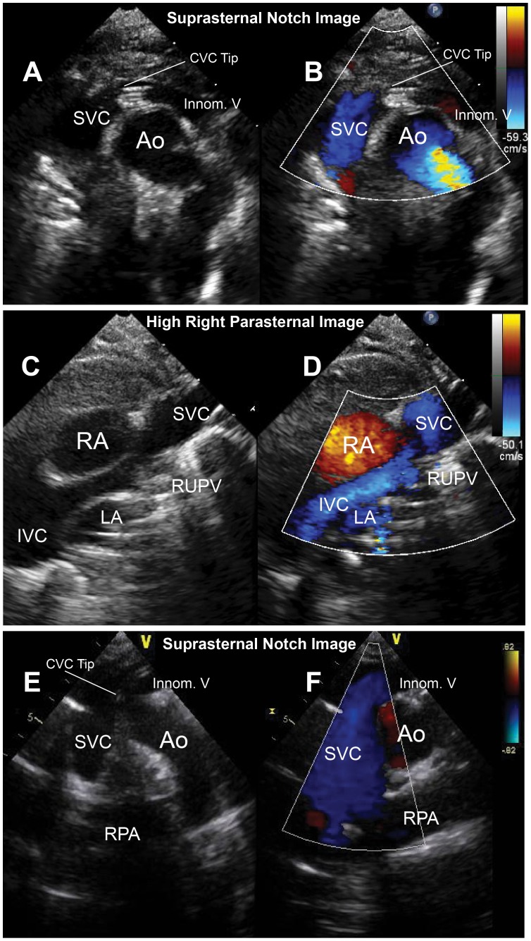
Figure 7. Representative images of superior vena cava and central venous catheter visualization in infants and children.
Panels A-D show suprasternal notch and high right parasternal 2-dimensional and color Doppler images of the superior vena cava in an infant. Arrows show the central venous catheter tip. Panels E and F demonstrate the superior vena cava and central venous catheter tip in a 14-year old child.

