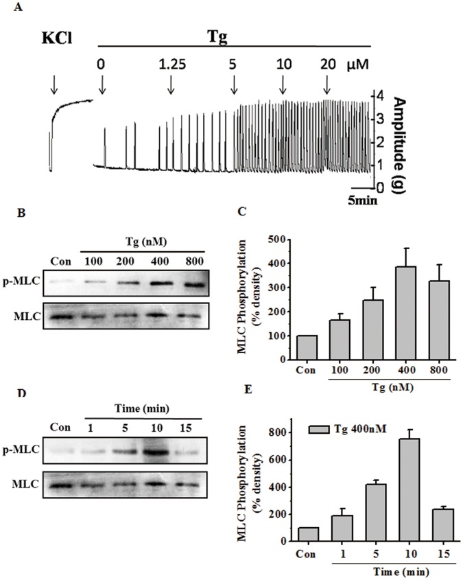
Figure 2. Tg dose- and time-dependently induced contractions of rat uterine strips and MLC20 phosphorylation in rat myometrial cells.
(A) Representative recording of rat myometrial contractions induced by cumulative doses of Tg. Muscle tension was recorded isometrically with a tension transducer connected to a polygraph system. The solution for each strip was first changed to 40 mM K+ for 10 min to ensure contractile viability and to determine maximum contraction. (B–E) Representative antibody reaction blots for the relative levels of MLC20 and pMLC20 in protein samples from rat myometrial cells treated with cumulative doses of Tg (B) or 400 nM Tg for different period of time (D). Quantitative analyses of the pMLC20-to-MLC20 ratio (C, E). Signal intensities for MLC20 and pMLC20 from three different blots were used for the quantitative analyses. Data are expressed as means ± SEM.

