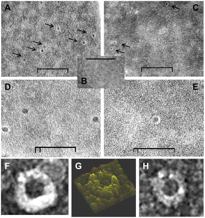
Figure 4. Ultrastructure of negatively stained RBC after exposure to cubozoan porins (Chironex fleckeri or Alatina moseri).
Transmission electron micrographs of 2% ammonium molybdate negative stained purified venom porin pretreated RBC showed the presence of distinct ring shaped pores. (A) Chironex fleckeri porin (A+B isoforms) treated human RBC membrane; (B) control mock treated RBC; (C) Alatina moseri porin exposed human RBC membrane; (D) higher magnification of A; (E) higher magnification of B; (F) and (H) highest magnification of individual exemplar pores from panel B; (G) 3-D modeling using analySIS EsiVision 3.2.0. Inner and outer diameter diameter of pores measured approximately 12 nm and 25 nm (size bars: 200 nm in panels A–C, 100 nm in panels D and E).

