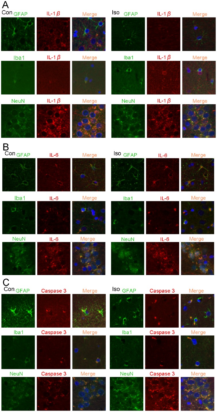
Figure 6. Expression of interleukin 1β (IL-1β), IL-6 and activated/cleaved caspase 3 in rat brain tissues.
Eighteen-month-old Fisher 344 rats were exposed to or were not exposed to 1.2% isoflurane for 2 h. Hippocampus was harvested at 48 h after isoflurane exposure for immunofluorescent staining of IL-1β (red), IL-6 (red), cleaved caspase 3 (red), NeuN (green), glial fibrillary acidic protein (GFAP, green) and ionized calcium binding adaptor molecule 1 (Iba1, green). The merged panels also include Hoechst staining (blue) to show cell nuclei. A: co-staining of IL-1β with GFAP, Iba1 and NeuN. B: co-staining of IL-6 with GFAP, Iba1 and NeuN. C: co-staining of cleaved caspase 3 with GFAP, Iba1 and NeuN. Con: control, Iso: isoflurane.

