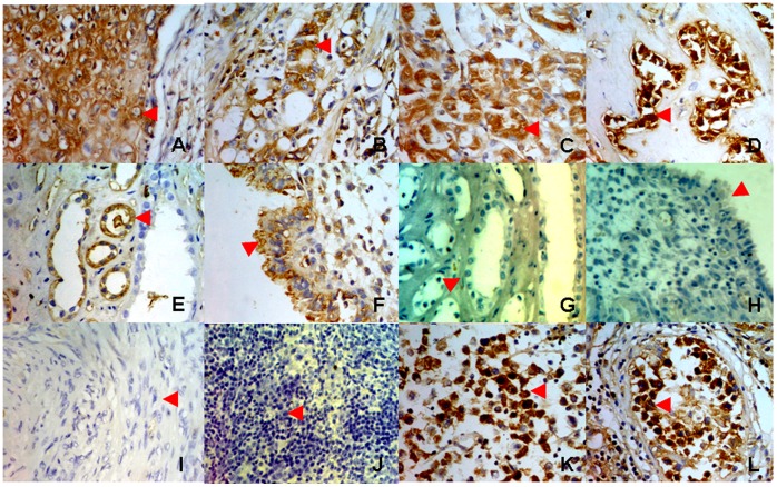
Figure 1. Detection of IgM expression by immunohistochemistry on tissue microarray.
A, lung cancer cells; B, breast cancer cells; C, liver cancer cells; D, pancreatic cancer cells; E, renal tubule epithelial cells; F, endometrium epithelial cells; G, renal tubule epithelial cells, stained with goat anti-mouse IgG-HRP only, as a negative control; H, endometrium epithelial cells, stained with goat anti-mouse IgG-HRP only, as a negative control; I, leiomyoma cells; J, T lymphoma cells; K, seminoma cells; L, spermatocytes.

