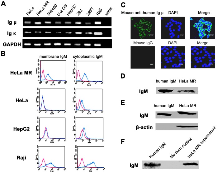
Figure 3. IgM expression in human non-B cell-derived cancer cell lines.
A, detection of Ig µ and Ig κ transcripts in multiple cancer cell lines by semi-nested RT-PCR. HeLa and HeLa MR, cervical cancer cell lines; SW480, colon cancer cell line; U-2 OS, osteosarcoma cell line; HepG2, hepatic cancer cell line; 293 and 293T, human embryonic kidney cell lines. Raji, human B lymphocytic leukemia cell line, as positive control; GAPDH, internal control. B, flow cytometry study using mouse anti-human Ig µ mAb showed that IgM was localized not only on the plasma membrane but also in the cytoplasm of epithelial cancer cells, especially HeLa MR cells. Red line, isotype control IgG1; Blue line, anti-human IgM. C, Confocal microscopy analysis of HeLa MR cells using mouse anti-human Ig µ mAb showed that IgM was present both on the cell membrane and in the cytoplasm. Mouse IgG, as isotype control. D, whole IgM was detected in HeLa MR cells by non-reducing SDS-PAGE (without β-mercaptoethanol) and Western blot with goat anti-human Ig µ polyclonal antibody. Human IgM, as positive control. E, IgM expression was detected in HeLa MR cells by reducing SDS-PAGE (with β-mercaptoethanol) and Western blot with mouse anti-human Ig µ mAb. Human IgM, as positive control; β-actin, internal control. F, IgM was also detected in the cultural supernatant of HeLa MR cells by reducing SDS-PAGE and Western blot using mouse anti-human Ig µ mAb. Human IgM, as positive control; Medium, as negative control.

