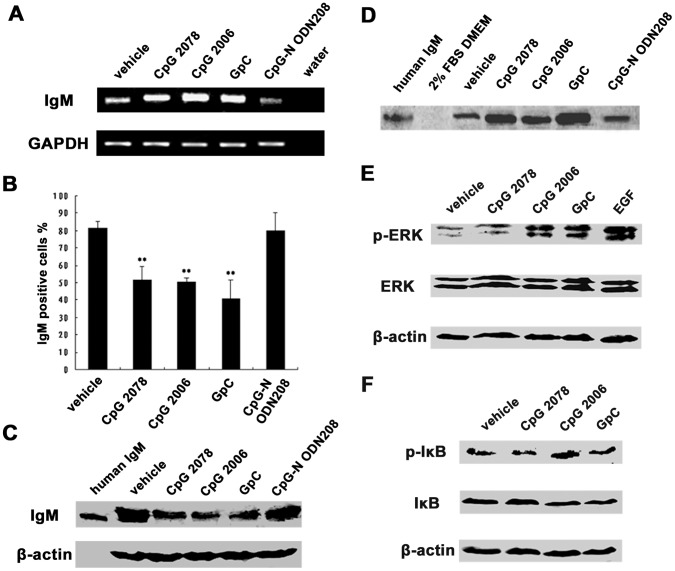
Figure 7. TLR9 agonists stimulated human epithelial cancer cells to secrete IgM.
A, Ig µ mRNA level was analyzed by semiquantitative RT-PCR after stimulation by CpG 2006, and the two non-CpG ODN controls, CpG 2078 and GpC. CpG-N ODN208, as negative control; GAPDH, internal control. B, flow cytometry analysis showed that cytoplasmic IgM was decreased after stimulation with CpG 2006, CpG 2078, and GpC. CpG-N ODN208, as negative control. **P<0.01 vs. vehicle control. C, Western blot analysis showed that cytoplasmic IgM was decreased after stimulation with CpG 2006, CpG 2078, and GpC. CpG-N ODN208, as negative control; β-actin, internal control. D, Western blot analysis showed increased level of secreted IgM after stimulation with CpG 2006, CpG 2078, and GpC. CpG-N ODN208, as negative control. E, phosphorylation of ERK, a molecule downstream of TLR9 signaling cascade, was detected by Western blot after stimulation with CpG 2006, CpG 2078, and GpC. EGF, epidermal growth factor was used as a positive control for ERK phosphorylation. F, Western blot analysis showed that IκB, a molecule in NF-κB pathway, also was phosphorylated after stimulation with CpG 2006, CpG, 2078 and GpC.

