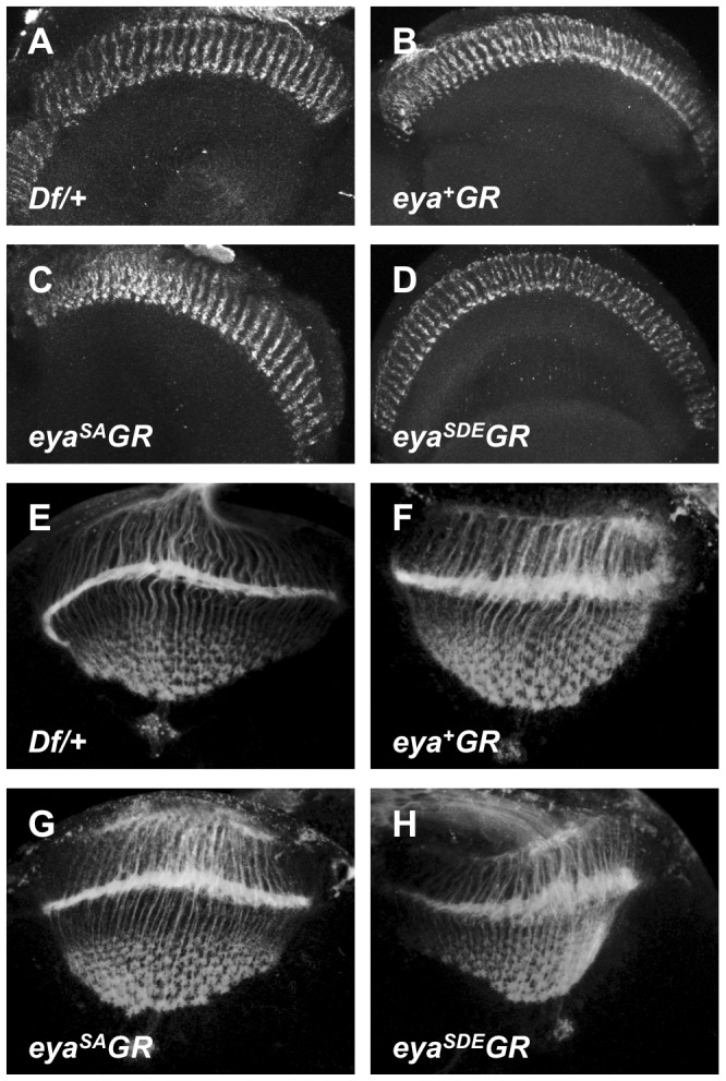
Figure 5. Photoreceptor axon projections are normal in flies rescued with eya*GR.

Photoreceptor axon projections to the optic lobe of Df/+ (A, E), eyacliIID/Df; eya+GR/+ (B, F), eyacliIID/Df; eyaSAGR/+ (C, G), and eyacliIID/Df; eyaSDEGR/+ (D, H) are indistinguishable from each other in both adults (A–D) and wandering third instar larvae (E–H). Axon projections are visualized with anti-Chaoptin (24B10).
