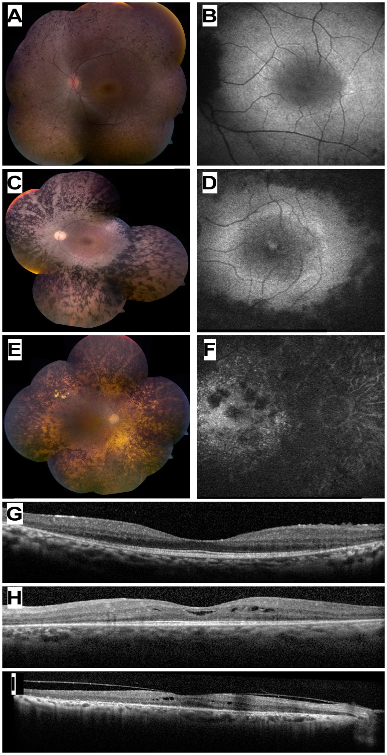
Figure 2. Ocular Phenotype of patients who are homozygous for the USH1C c.1220delG mutation.
(A, C, E) Color fundus photographs of three patients aged 13 (MOL0486 II:3), 33 (TB16-R12), and 72 (MOL1023-1) years old, respectively. Note the increasing severity of fundus changes with age, and the presence of dense bone spicule-like pigmentation. (B, D, F) Corresponding fundus autofluorescence imaging of the macular area in the three patients shown in A,C,E. Note hyeprfluorescent rings around the foveas in the younger patients, and hypofluorescent areas of atrophy that encroach upon the macula in the 33 year-old patient and invade the macula in the 72 year-old. (G, H, I) Horizontal optical coherence tomography (OCT) cross-sections through the fovea in the 13 yo (panel G), 33 yo (H) and 72 yo (I) USH1C patients showing progressive loss of retinal and particularly photoreceptor layer thickness with age. Intra-retinal cysts of fluid (cystoid macular edema) are evident in two of the cases (H, I).

