
Figure 1. Confocal microscopy.
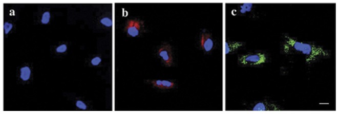
Unlabeled control (A), GadofluorineM-Cy-labeled hMSCs (B) and anti-dextran-FITC stain of Ferucarbotran-labeled hMSCs (C). All cells have been counterstained with DAPI (blue). Note the cytoplasmatic localization of both contrast agents (B, C) whereas no contrast agent could be seen in the nucleus. Scale bar = 10 µm.