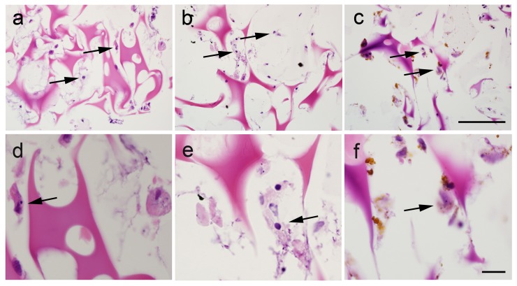
Figure 7. Histology of hMSC implants (H&E staining).
All scaffolds show a homogeneous pink staining of the agarose matrix within the gelatin sponge. Injected hMSCs (arrows) can be seen after injection of unlabeled cells (A and D), GadofluorineM-Cy-labeled cells (B and E) and Ferucarbotran-labeled cells (C and F). While iron oxides can be delineated, GadofluorineM-Cy remains invisible at higher magnification and light microscopy (Scale bar = 100 µm).

