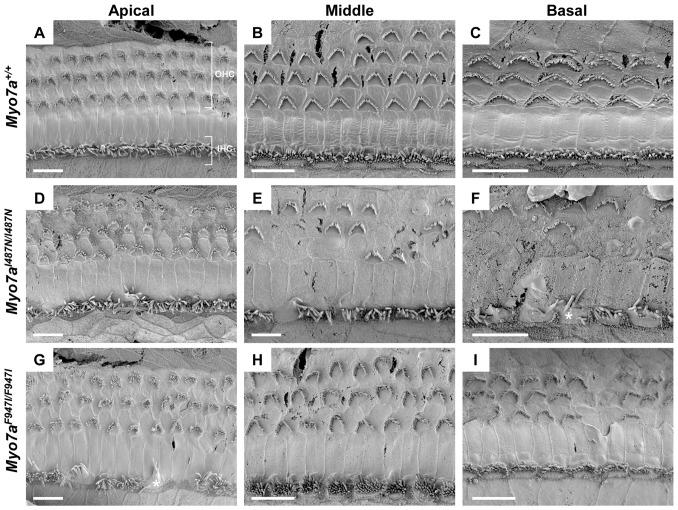
Figure 5. Scanning electron micrographs of cochlear sensory epithelium from Myo7a mutant strains at 8 weeks old.
(A–C) Myo7a+/+, (D–F) Myo7aI487N/I487N ewaso and (G–I) Myo7aF947I/F947I dumbo mice at the apical, middle and basal cochlear level. Signs of degeneration and/or mis-orientation of OHC bundles is evident in both Myo7aI487N/I487N ewaso (D–F) and Myo7aF947I/F947I dumbo (G–I) mice at all levels of the cochlea. This appears to be more severe in Myo7aI487N/I487N ewaso mutants, where many bundles are missing in the mid and basal regions (E and F). IHC bundles are also affected, appearing disorganised and/or showing signs of fusion in the basal levels of Myo7aI487N/I487N ewaso cochleae (asterisk in F), and conversely in the apical region of Myo7aF947I/F947I dumbo mutants (asterisk in G). Scale bar; 10 µM (A–I).

