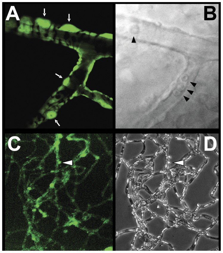
Figure 1. Appearance of retinal capillaries from transgenic mice.
(A) Confocal microscopy of a retinal capillary from an SMAA-GFP mouse illustrating the appearance of pericytes with their finger-like cytoplasmic projections compassing the vascular endothelial cells. GFP is present in the cytoplasm of the pericytes and their processes (white arrows). (B) Bright field image of the same capillary showing the presence of red blood cells (arrowheads). (C) Appearance of retinal capillaries isolated by trypsin digestion illustrating that GFP is retained in the intact pericytes and can be used for their identification. (D) Appearance of the same capillaries by light microscopy.

