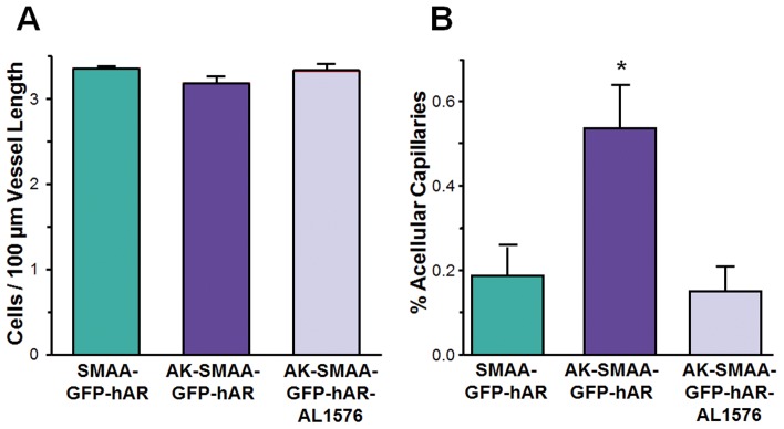
Figure 7. Changes in isolated retinal capillary capillaries from 18 week diabetic AK-SMAA-GFP mice and AK-SMAA-GFP-hAR mice treated with/without ARI.
In A the capillary cell density expressed as capillary nuclei/100 µm of capillary length is presented. In B the percent of acellular capillaries present in the examined neural retinal capillaries is presented. n = 5–7; mean ± S.E.M. * p≤0.03.

