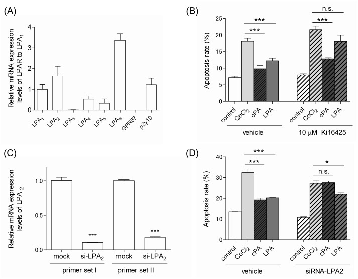
Figure 4. Effects of LPA receptors on the neuroprotective functions of cPA and LPA against CoCl2-induced apoptosis.
(A) Expression of LPA receptors in Neuro2A cells. Total RNA was extracted from Neuro2A cells, and the expression level of each LPA receptor was determined by quantitative real-time PCR. The expression levels were normalized to those of LPA1 and expressed in terms of the mean ± SE values. (B) The effects of Ki16425 on the neuroprotective functions of cPA and LPA against CoCl2-induced apoptosis of Neuro2A cells. Neuro2A cells were pretreated with or without 10 µM Ki16425 for 20 min. Subsequently, the cells were incubated with 300 µM CoCl2 in the presence of 10 µM cPA or LPA for 24 hours. The cells were then stained with FITC-Annexin V and subjected to flow cytometric analysis. The data represent the mean ± SE values from triplicate independent experiments (***P<0.001 vs. the CoCl2-treated group). (C) Expression of LPA2 in Neuro2A cells. Neuro2A cells were transfected with siRNA against LPA2 or non-target siRNA. Total RNA was extracted from each transfected Neuro2A cell, and the expression level of each LPA receptor was determined by quantitative real-time PCR. The expression levels of LPA2 was normalized to those of Neuro2A cells transfected with non-target siRNA. The resulting data represent the mean ± SE values (***P<0.001 vs. the mock group). (D) The effects of LPA2 knockdown on the neuroprotective effects of cPA and LPA against CoCl2-induced apoptosis of Neuro2A cells. Neuro2A cells transfected with either siRNA against LPA2 or non-target siRNA were incubated with 300 µM CoCl2 in the presence of 10 µM cPA or LPA for 24 hours. Cells stained with FITC-Annexin V were subjected to flow cytometric analysis. The data represent the mean ± SE values from triplicate independent experiments (*P<0.05, ***P<0.001 vs. the CoCl2-treated group; n.s., not significant).

