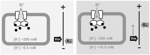
FIGURE 7. A hypothetic model to illustrate the cause of the K+-sensitive phenotype by the cAMP-gated MmaK in the host E. coli cell.
MmaK in the live E. coli is constitutively activated by the intracellular cAMP (black dots). Whether there is a K+ flux through MmaK depends on the membrane potential (Vm) and the equilibrium potential of K+ (EK). The Vm of an aerobically growing E. coli cell is ~ −130 to −150 mV, which is set by active proton extrusion through the respiratory components on the cell membrane (34) Left, when the external [K+] is low (~0.5 mM K+ in the tryptone medium), the EK is deep (~−155 mV, assuming [K+]in = 200 mM). The driving force for K+ (ΔμK) would be slightly positive to cause K+ efflux through MmaK and may result in a small hyperpolarization of the Vm. Right, adding 10 mM K+ to the medium increases EK (to ~−75 mV) and decreases the ΔμK (to ~−55 to −75 mV). The sizable negative ΔμK+ would drive K+ flux into the cell through the active MmaK and result in significant depolarization of the Vm toward EK. Because Vm is the major component of the protonmotive force (Δp), the reduced Δp may not be able to provide enough driving force for other cellular processes, and thus the cell growth stops. The lower final density in liquid and thinner colonies on semi-solid plates suggests that the MmaK-bearing cells are under stress even in tryptone medium (Fig. 3), consistent with there being a small, albeit tolerable, ΔμK even in growing cultures from the constitutively active MmaK channels.

