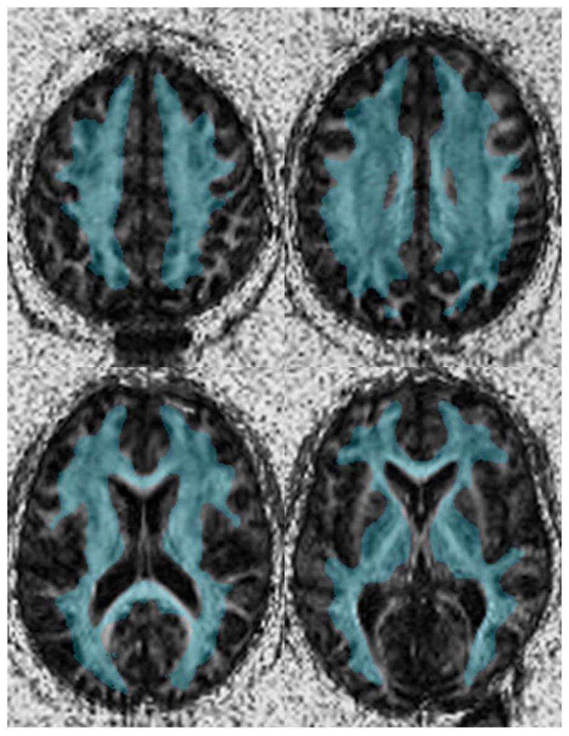
Figure 1.

The binary white matter mask used in the voxel based analysis overlaid on a single patient after spatial normalization. This image demonstrates that while this mask was created from the average FA image from all patients, the majority of the white matter is identified on each individual subject.
