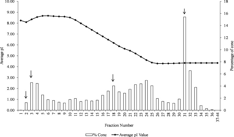
Fig. 2.
Elution profile and percentage concentration of SWA into various fractions during chromatofocusing. Chromatofocusing was performed using the ProteomeLab PF 2D protein separation system. The pH gradient was formed using two proprietary buffers: “ProteoSep Start” buffer at a pH 8.5 and “ProteoSep Elution” buffer at a pH 4.0. As shown, the SWA was well fractionated with fraction 31 containing most of the void proteins. Proteins identified were obtained from fractions numbers indicated by arrows (2, 3, 18 and 3).

