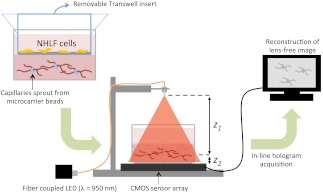
Fig. 1.
Schematic of our tissue culture system and lens-free computational imaging apparatus. NHLF fibroblasts are cultured on the top surface of a Transwell insert, which is placed within a 35 mm glass-bottom Petri dish. HUVECs were first cultured onto microcarrier beads, which were then embedded within a crosslinked fibrin hydrogel. Soluble signals released from the fibroblasts stimulate the HUVECs to spontaneously form capillaries. The insert was removed prior to imaging. A partially coherent fiber-coupled LED light source illuminates the Petri dish, which is placed above the CMOS chip to record the holographic image of the sample over a large field-of-view and extended depth-of-field; is the distance between the light source and the object; is the distance between the objects (capillaries) and the active area of the detector, which is changed by placing or removing a glass coverslip underneath the sample. A computer reconstructs lens-free images according to described algorithms.

