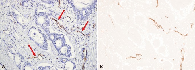
Fig. 3.
Morphometric study of microvessels. Microvessels were immunostained using a CD31-related antigen-specific mouse monoclonal antiboy. (A) A photomicrograph (200×) of a hot spot in a representative tumor section shows immunostained microvessels (arrows) in brown. (B) Microvessels are highlighted by a color threshold setting to distinguish the objects of positive staining from the counter-stained background tissue.

