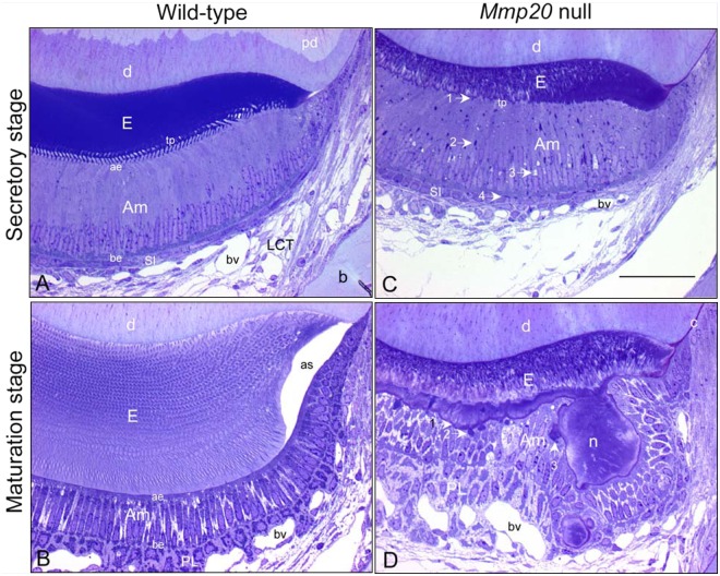
Figure 1.
Semi-thin (0.5 µm) sections from glutaraldehyde-fixed, decalcified, and plastic-embedded mandibular incisors of wild-type (A,B) and Mmp20 null (C,D) mice stained with toluidine blue to illustrate the appearance of enamel organ cells at mid-secretory stage (A,C) and near-mid-maturation stage (B, D) of enamel development; magnification bar in C equals 50 µm and applies to all panels. The absence of MMP20 does not alter the basic configuration of the enamel organ in terms of size, number, types and arrangement of cell layers (compare C with A, D with B), the tall columnar appearance of ameloblasts as they form enamel (compare C with A, Am), and their shorter and wider appearance as they participate in enamel maturation (compare D with B, Am). However, there are several location-specific alterations in the enamel organ of Mmp20 null mice, including: (a) thinner, highly disorganized, and poorly formed inner and outer enamel layers (E) (compare C with A, D with B); (b) abnormalities in secretory-stage ameloblasts, such as disruption of row organization of Tomes’ processes (tp) (compare C with A), irregular and ragged apical surface applied to forming enamel (C, arrow 1), presence of large dense-staining bodies (C, arrow 2) and vacuoles (C, arrow 3) in supranuclear region of ameloblasts and dense-staining bodies at their bases near stratum intermedium cells (SI) (C, arrow 4); and (c) abnormalities in maturation-stage ameloblasts, including undulating and metachromatic (mauve)-staining at the enamel surface (D, arrow 1), presence of large dense-staining bodies in apical region of modulating ameloblasts (D, arrow 2), some appearing in continuity to the outer edges of matrix nodules (n) projecting from the enamel surface (D, arrow 3) that are covered by unevenly distributed and distorted ameloblasts (Am) and papillary layer cells (PL, D). Other abbreviations: pd, predentin; D, dentin; ae, apical end; be, basal end; bv, blood vessel; as, artifact space; b, bone; c, cementum.

