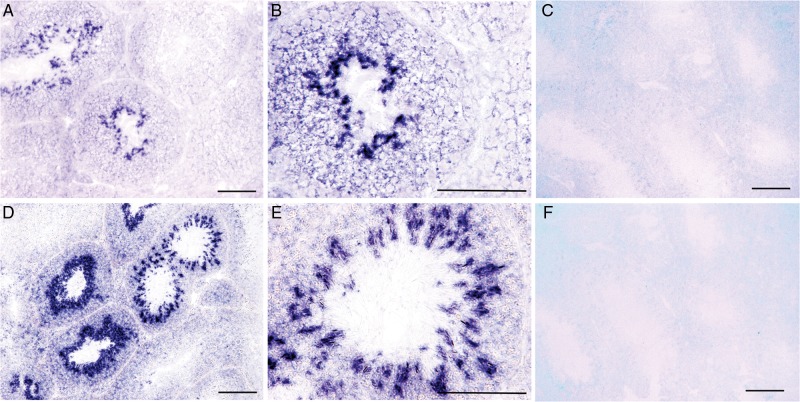
Figure 2.
In situ hybridization of mouse testicular sections with Tas2r probes. (A and D) Antisense probes of Tas2r105 (A) and Tas2r108 (D) hybridized to subsets of cells in some seminiferous sections (blue staining). (B and E) High-magnification images of Tas2r105 (B) and Tas2r108 (E) show that the stained cells were post-meiotic cells. (C and F) No signals with the sense probes Tas2r105 (C) and Tas2r108 (F) were detected. Scale bars: 100 μm.

