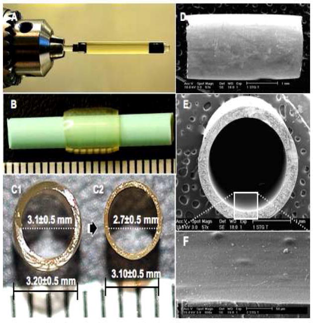
Fig. 2. Fabrication of gelatin tubes.

(A) A glass tube (ID; 3.2±0.5mm, OD; 5.0mm) containing a glass rod (ID; 3.1±0.5mm), both siliconized, were supported horizontally by a rotator stirrer (1000 rpm) and irradiated using blue visible light. (B) The resulting transparent gelatin tube (wall thickness, 500μm) was then reinserted (see text) on a thinner Teflon rod (OD; 2.7±0.5mm). The tube diameter before (C1) and after (C2) dehydration is shown. Longitudinal view of the tube outer surface (D), a cross-sectional view (E), and the inner surface (F) are illustrated in scanning electron micrographs.
