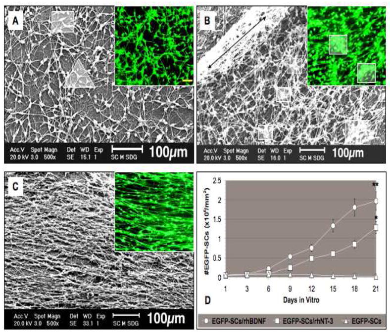
Fig. 6. Scanning electron micrographs of EGFP-SCs on gelatin membranes 21 days in vitro.

(A) EGFP-SCs seeded on membranes without neurotrophins were typically bipolar (square in A) but were not aligned; tri-polar cells (triangle in A) were sometimes observed in the SEM. Green flourescent cells are shown in the smaller panels. (B) rhNT-3 membranes contain high numbers of EGFP-SCs strongly attached to their surfaces, forming a compact network sometimes piled up in layers (long black double arrowhead) with some clumping of cells (white squares in smaller panels). (C) Membranes containing rhBDNF and EGFP-SCs supported a highly aligned cell array. Mean numbers of EGFP-SCs in the three groups are presented in panel D. Error bars represent means ± SD (n = 6 per group). Significant statistical differences were accepted at *p < 0.05 and **p < 0.01 compared to control cultures. Scale bar; 100μm in fluorescence photographs.
