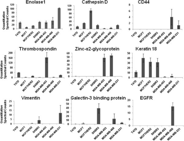
Figure 1.
Quantitative comparison of proteins detected by LC-MS/MS in the proximal fluid of seven breast cancer cell lines. In triplicate, MS/MS data of proteins identified in the proximal fluid from each breast cancer cell line was quantitatively determined by spectral counting in Scaffold software. Enolase 1, cathepsin D, CD44,Thrombospondin 1, zinc-α2-glycoprotein, keratin 18, vimentin, galectin-3 binding protein, and EGFR were chosen to represent HER2 negative and hormone receptor positive cell lines T47D and MCF-7, HER2 positive SKBR-3 and MDA-MB-453, triple negative breast cancer MDA-MB-468 and MDA-MB-231, and hormone receptor positive MCF-7 transfected with HER2 (MCF-7HER2).

