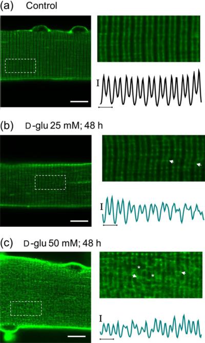
Figure 7.
Transverse tubular system disruption accompanying d-glucose exposure. Confocal images of FDB muscle fibers maintained in isotonic control (5.56 mmol/L d-glucose) medium (a) (n = 22 fibers, three mice), 25 mmol/L d-glucose (b) (n = 36 fibers, three mice) and 50 mmol/L (c) (n = 28 fibers, three mice) for 48 h and then stained with di-8-ANEPPS (see protocol in Figure 9a). Scale bars: 10 μm. (a–c) Right panels, zoom-in versions of boxed regions indicated in left panels. Traces below zoom-in images are averaged fluorescence profiles across the box, vertical scale bar: 500 A.U.; horizontal scale bar: 2 μm. Di-8-ANEPPS staining reveals that the normally regular transverse-tubule morphology (a) is moderately affected in fibers exposed to 25 mmol/L d-glucose for 48 h (b), but is remarkably disrupted in fibers exposed to 50 mmol/L d-glucose for 48 h (c). The asterisks indicate places of transverse tubule dilation and the arrows indicate locations where adjacent transverse tubule appear to make close contact. FDB, flexor digitorum brevis; NFAT5, nuclear factor of activated T-cells 5; A.U., arbitrary units. (A color version of this figure is available in the online journal)

