
Figure 1.
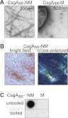
Amyloid-like aggregates formed by secreted CsgAss-NM. (A) Electron micrographs of immunolabeled CsgAss-NM (left) and CsgAss-M (right) scraped cell samples. Fibrils were detected only with the CsgAss-NM sample. (B) Micrographs of CsgAss-NM scraped cell sample harvested from CR-containing agar. Extracellular material binds CR (left) and displays apple-green birefringence when viewed between crossed polarizers (right). (C) CsgAss-NM, but not CsgAss-M, scraped cell samples contain SDS-resistant aggregates that are solubilized upon boiling, as detected by filter retention using an antibody that recognizes the M domain.