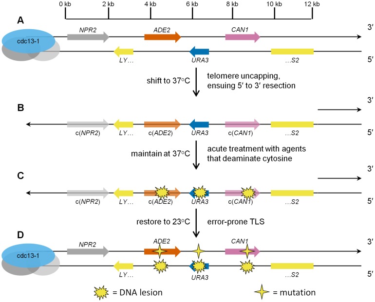
Figure 1. Schematic representation of ssDNA mutagenesis reporter system.
(A) Three reporter genes, ADE2, URA3, and CAN1, were relocated from their respective native genomic loci into the subtelomeric region of Chromosome V, within a haploid budding yeast strain bearing the temperature-sensitive cdc13-1 mutation, thus creating strain ySR127. The 0 kb mark in the scale bar denotes the start of unique DNA sequence (conversely, the end of telomeric repeat sequences). (B) Shifting ySR127 cells to 37°C results in telomere uncapping. Subsequent 5′ to 3′ resection results in a long 3′ ssDNA overhang. c(ADE2) and c(CAN1) denote the complement of the two genes. (C) Cells then undergo acute treatment with agents that deaminate cytosine, e.g. human APOBEC3G or sodium bisulfite, which induce lesions in the 3′ ssDNA overhang. (D) Shifting back to permissive temperature (23°C) restores the subtelomeric DNA to double-stranded form. Error-prone bypass of lesions formed in ssDNA generates a strand-coordinated, multi-mutation signature, which is detected by simultaneous loss of function in two or more of the reporter genes, and verified by sequencing of individual multi-loss-of-function isolates.

