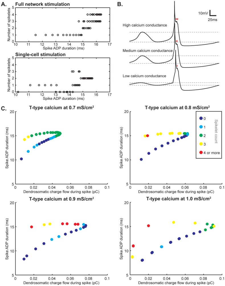
Figure 3. Factors underlying AP spikelet count.
A. Correlation between spike ADP duration and AP spikelet count. In general, a longer ADP increases the chances of larger amounts of spikelets on top of the somatic ADP. This is most readily apparent when stimulating the entire network (top panel). Still, the ADP duration by itself does not provide an adequate explanation for the amount of spikelets that are part of the spike shape, since the number of spikelets can still vary considerably even within a millisecond bin both when the entire networks fires and when only one cell does. When only one cell in the network fires, the relation between ADP duration and number of spikelets does not appear to be linear, even though on average a higher number of spikelets is still more likely at longer ADP durations (bottom panel). B. Changing the intrinsic conductance values of the low-threshold calcium current changes the amplitude of the oscillations (as indicated by the dashed lines aligned with the peaks and troughs of the depicted traces), but also the number of spikelets (time of occurrence is indicated with red markers in each depicted trace). The number of spikelets decreases as the T-type calcium expression level decreases. C. Spike ADP and dendrosomatic coupling currents as AP spikelet count determinants. Single cells in a 9-cell network were stimulated for all of the simulation results shown. Warmer colors represent higher numbers of AP spikelets (color-coding is the same for all four panels and indicated in the figure). Spike ADP duration and dendrosomatic charge flow form a trajectory. It is readily apparent from all four panels that higher numbers of spikelets are more likely to occur at longer ADPs, but in addition decreased charge flow increases the chance of generating an AP with more spikelets. As a result, the prediction of AP spikelet count can be improved when taking both ADP duration and dendrosomatic charge flow into account. At different T-type calcium expression levels, the range of possible spikelet counts and the distributions thereof vary. Clearly, spike ADP and dendrosomatic coupling currents are major determinants for the AP spikelet count measured at the soma, but other currents both intra- and intercellular can cause local phenomena in the distributions along the trajectories shown.

