FIGURE 3:
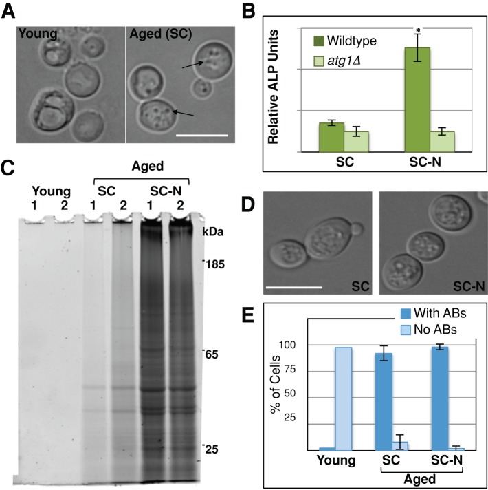
Accumulation of SDS-insoluble protein correlates with the presence of autophagic bodies. (A) Young and aged cells were harvested as described, and vacuolar morphology was evaluated for the presence of ABs (arrows) by light microscopy. (B) After 48 h of postmitotic growth in synthetic complete media (SC), wild-type (TN121) or atg1Δ (TN123) cells carrying the Pho8Δ60 reporter were starved for nitrogen (SC-N) for 8 h, and alkaline phosphatase (ALP) activity was measured (n = 4). (C–E) Wild-type cells were grown into the postmitotic state and harvested (young) or aged in SC or SC-N media for 48 h. Cells were harvested, and SDS-insoluble protein extracted from 4 × 10⁷ cells was analyzed by SDS–PAGE (C) or the percentage of cells with ABs quantitated (D, E). More than 200 cells were quantitated for each replicate (n = 3). White bars, 10 μm. *p < 0.01.

