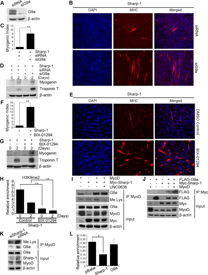
FIGURE 3:
Inhibition of G9a rescues Sharp-1–imposed differentiation block. (A) C2C12 cells overexpressing Sharp-1 were transfected with control siRNA or siG9a. G9a knockdown was determined by Western blot. (B) Cells were induced to differentiate and stained with anti-MHC antibody. Nuclei were stained with DAPI. (C) Differentiation was quantified by calculating myogenic index. (D) Myogenin and troponin T expression was determined by Western blot. (E–G) Sharp-1–overexpressing cells were incubated with DMSO (vehicle) or BIX-01294. Differentiation was assessed using anti-MHC antibody (E), myogenic index (F), and expression of myogenin and troponin T by Western blot (G). (H) Sharp-1–overexpressing cells were treated with DMSO or BIX-01294 for 0 and 2 d. ChIP assays were done using anti-H3K9me2 antibody on the myogenin promoter. (I) C2C12 cells were transfected with Myc–Sharp-1 and MyoD. MyoD was immunoprecipitated and analyzed for association with G9a and methylation in the absence and presence of UNC0638 treatment using anti-G9a and anti-Me Lys antibodies, respectively. Lysates were analyzed for expression of G9a, MyoD, and Sharp-1. (J) C2C12 cells were transfected with Myc–Sharp-1 and MyoD and G9a as indicated. The association of Sharp-1 with G9a was analyzed in the absence and presence of MyoD. (K) Endogenous MyoD methylation and association with G9a was examined by immunoprecipitation from siSharp-1 and siRNA cells using anti-Me Lys and anti-G9a antibodies. Input shows expression of G9a, Sharp-1, and MyoD in lysates. (L) MyoD occupancy on the myogenin promoter was analyzed by ChIP assays in myoblasts overexpressing pBabe, Sharp-1, or G9a. Error bars indicate mean ± SD.

