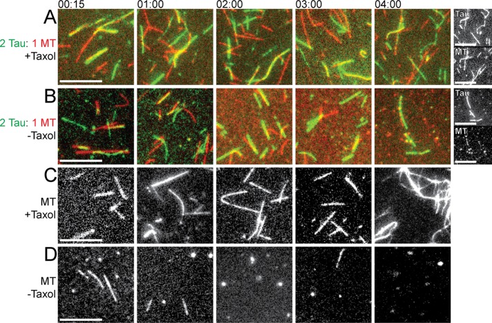
FIGURE 3:
Tau filaments form in the presence of MTs and remain after MTs dissociate. MTs (4 µM) were incubated at 37°C in the absence or presence of Tau (2 µM) and/or the MT-stabilizing drug Taxol (10 µM). Samples of the incubations were taken at different time intervals (from 15 min to 4 h, as indicated) and imaged. Colored images display tubulin as red and Tau as green and their overlay as yellow. Scale bars: 8 µm. (A) MTs incubated with Taxol and Tau; the far right pair of panels shows grayscale images for the 4-h time point. (B) MTs incubated with Tau in the absence of Taxol; the far right pair of panels shows grayscale images for the 4-h time point. (C) MTs incubated alone with Taxol. (D) MTs incubated without Taxol or Tau. Note that MTs without Taxol dissociate over time (more slowly when Tau is present), but the Tau filaments remain. Images shown represent the typical results of more than six different fields of collected data.

