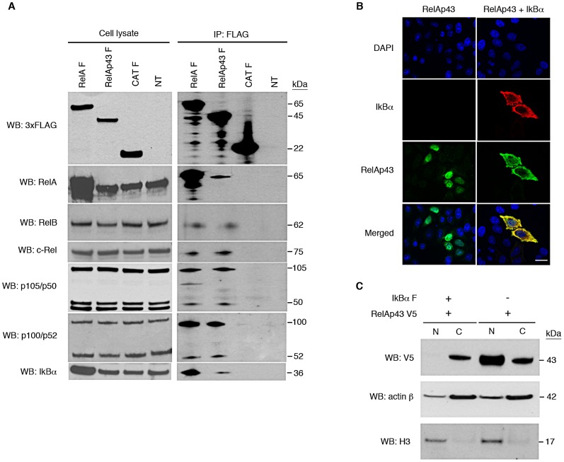
Figure 2. RelAp43 interacts with all human members of the NF-κB family and is supported by IκBα.
All experiments were performed three times independently. (A) IP using anti-FLAG antibody of FLAG-tagged RelAp43 (RelAp43 F) or RelA (RelA F) or CAT (CAT F) expressing cells or non-transfected cells (NT). The presence of FLAG-tagged protein or endogenous transcription factors of the NF-κB family was analyzed by western blot using specific antibodies in cell lysates either before (left panel) or after IP (right panel). (B) Cellular localization of transfected RelAp43 (green) analyzed by immunofluorescence and apotome imaging in absence (left panel), or in presence of transfected IκBα (red, right panel). Nuclei were visualized using DAPI staining. The scale bar corresponds to 2 µm. (C) Western Blot analysis of nuclear (N) and cytosolic fractions (C) of RelAp43 V5 and IκBα F co-transfected cells after nuclear-cytosolic fractionation. Nuclear and cytosolic fractions were controlled using anti histone H3 and anti actin β antibodies, respectively. See also Figure S2.

