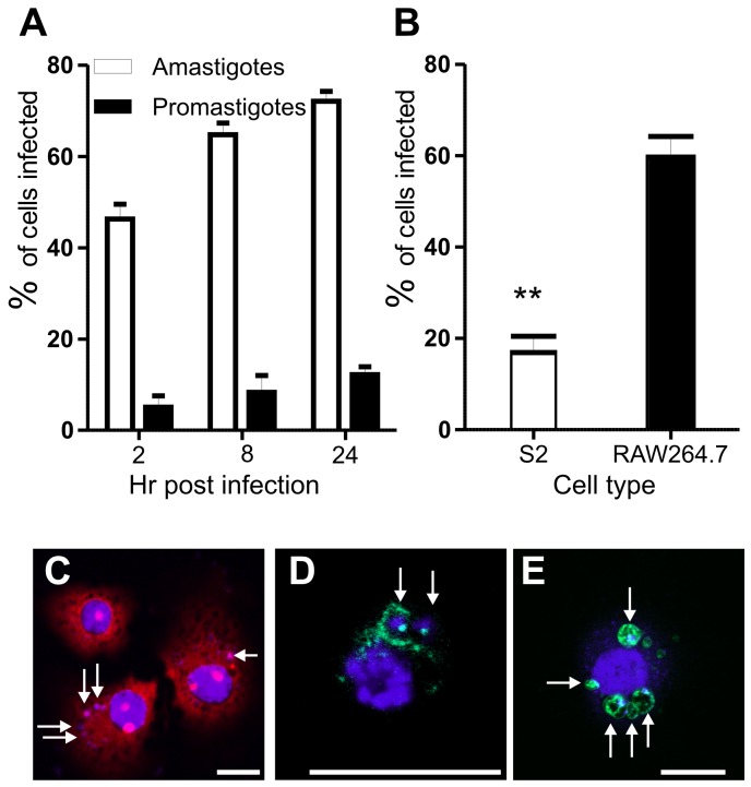
Figure 1. Infection of S2 cells with different Leishmania species and life cycle stages.
A. Percentage of S2 cells infected after incubation with either L. donovani amastigotes or L. major late stationary phase promastigotes over a 24 hr time course. B. Comparison of infection levels of S2 cells and mammalian RAW264.7 macrophages incubated with late stationary phase L. major promastigotes. p = 0.0025; error bars in A and B indicate one standard error (SE). C. S2 cells infected with L. donovani amastigotes for 24 hr are stained with 1 µg/ml DAPI, 20 µg/ml propidium iodide; parasites were pre-labelled with 10 µM CFSE. D. S2 cells infected with L. donovani amastigotes (24 hr post infection) stained with anti-ARL8, a late endosomal/lysosomal marker. E. S2 cells expressing LAMP-GFP (green) infected with L. donovani amastigotes for 12 hr. C – E, scale bars, 10 µm. Arrows indicate intracellular parasites.

