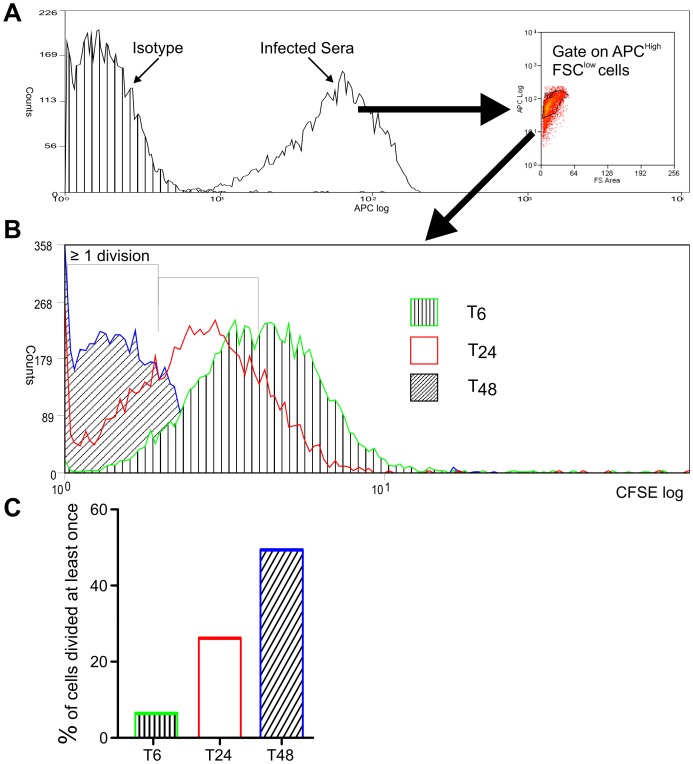
Figure 3. Replication of L. donovani amastigotes within S2 cells.
S2 cells were infected with CFSE-labelled parasites for 6 hr, followed by washing to remove external parasites. At the time points indicated, S2 cells were lysed with saponin to release amastigotes. A. Parasites were identified from cell debris by staining with the sera from an infected hamster conjugated to fluorescent allophycocyanin (APC), followed by flow cytometry and gating on APC high, forward scatter low cells. B. Flow cytometry overlay of CFSE staining of parasites identified in A, released after 6, 24 and 48 hr. C. By gating on CFSE staining in B, the percentage of parasites that had replicated at least once was calculated at each time point. Data are representative of two independent experiments.

