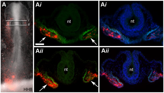
Figure 3. Descendants of cells from region B are located within the splanchnic mesoderm and express Isl1.
(A) Bright field and fluorescent image showing the location of fluorescently labelled descendants of cells from region B at stage HH8 (red cells). (A i –A ii) Sections through embryo in (A) were stained for Isl1 protein expression (green). DAPI signal (blue) has been superimposed to provide a clearer picture of the morphology of the tissue. White arrows in (A i –A ii) point to the splanchnic mesoderm where fluorescent cells (DiI intercalated within the cell membrane) that express Isl1 are located. Neural tube (nt), (n = 11). Scale bar = 50 µm.

