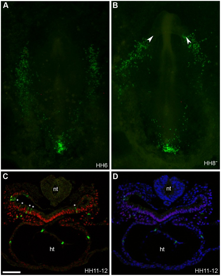
Figure 6. CAG-GFP cells grafted within region B are located in the splanchnic mesoderm and heart.
(A–B) Bright field and fluorescent images of embryos at stage HH6 (A) and HH8- (B) showing location of CAG-GFP labelled cells (green cells) which were grafted within region B of the primitive streak at stage HH3 though to HH3+. White arrow heads in (B) mark the location of the intestinal portals. (C) Section of grafted embryo stained for Isl1 protein expression (red). (D) DAPI signal (blue) has been superimposed to provide a clearer picture of the morphology of the tissue. At stage HH11-12 CAG-GFP labelled cells grafted to region B were located within the pharyngeal endoderm (cells marked with white asterix), splanchnic mesoderm and the heart (ht). Images were taken on confocal microscope. Neural tube (nt), (n = 19). Scale bar = 60 µm.

