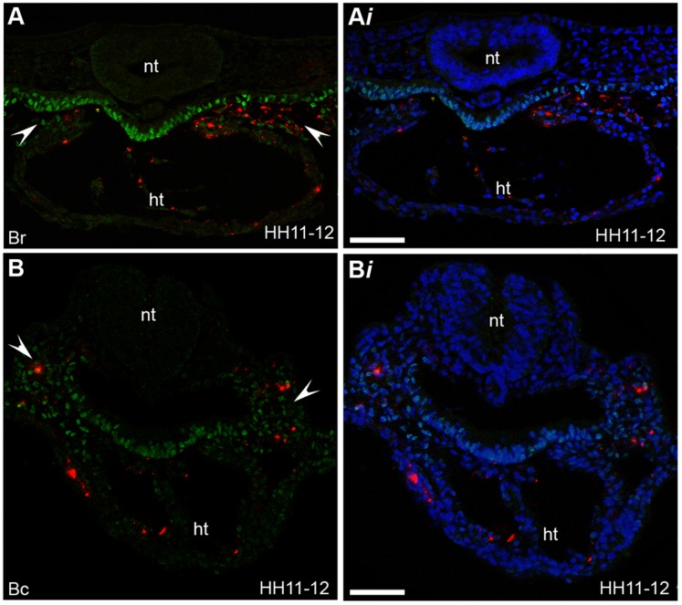
Figure 7. Region B is the ingression site of CPCs located within the splanchnic mesoderm and heart.
(A–B i) Sections of labelled embryos at stage HH11-12 stained for Isl1 protein expression (green). (A) Fluorescent descendants of cells (red cells) labelled in rostral subdivisions within region B (Br) are located within the splanchnic mesoderm (indicated with white arrow heads) and the heart (ht). (B) Fluorescent descendants of cells (red cells) labelled in caudal subdivisions within region B (Bc) are located within the splanchnic mesoderm (indicated with white arrow heads) and the heart (ht). (Ai, Bi) DAPI signal (blue) has been superimposed to provide a clearer picture of the morphology of the tissue. Images taken on confocal microscope, neural tube (nt) (n = 31). Scale bars = 60 µm.

