
Figure 2. Real-time visualization of sister fork uncoupling during unwinding of doubly tethered DNA.
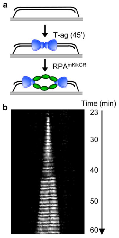
a, T-ag was drawn into a flow cell containing doubly tethered λori. After 45 min, RPAmKikGR was introduced and mKikGR was imaged for 60 min. b, Kymograph of mKikGR fluorescence. Minutes denote time after introduction of RPAmKikGR.