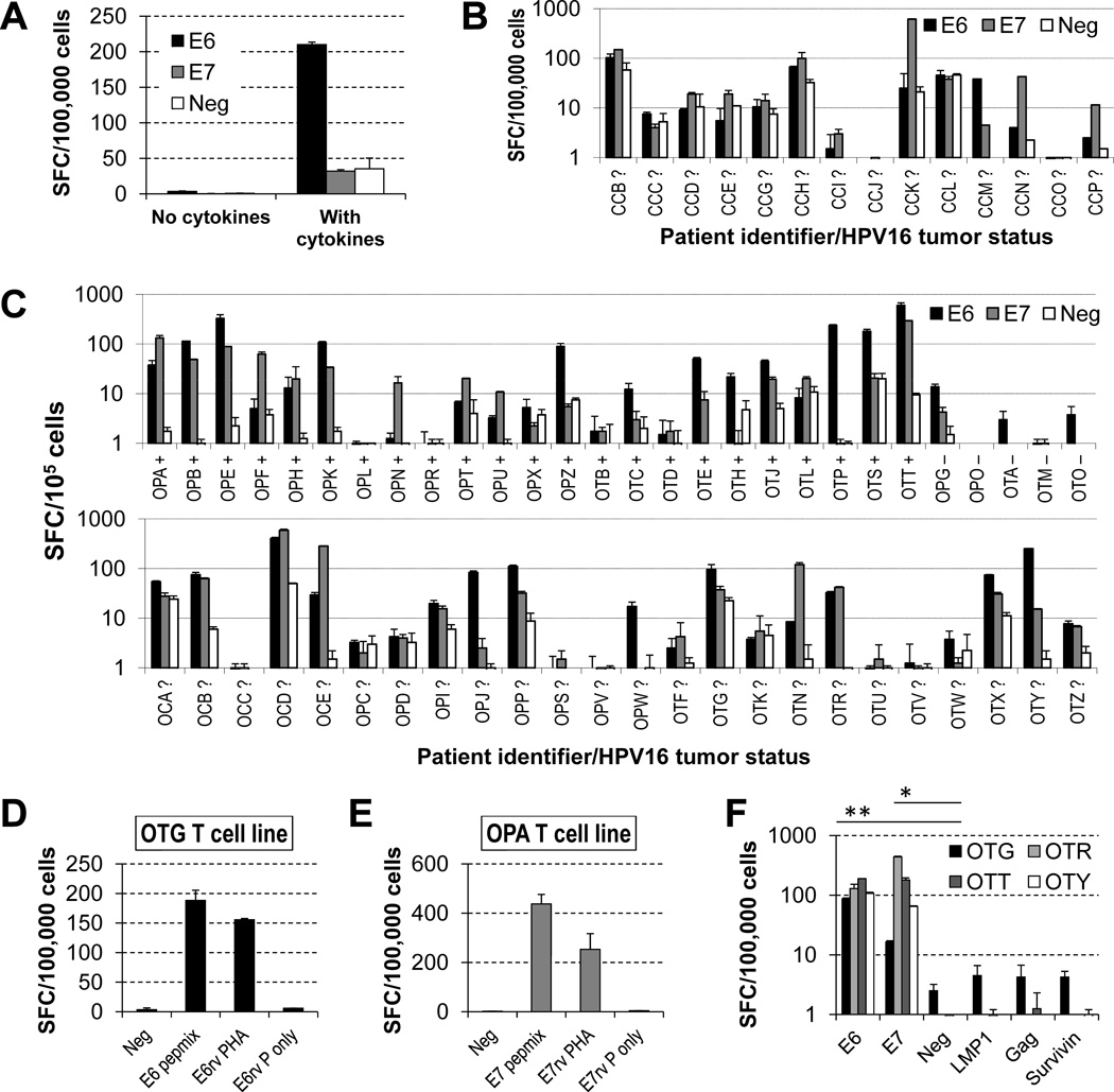
Figure 1. HPV16 E6/7-specific T cells can be expanded from the peripheral blood of patients with HPV16-associated cancers using autologous DCs.
Patients PBMCs were stimulated twice with autologous DCs loaded with E6 and E7 pepmixes, with or without accessory cytokines (IL-6, -7, -12 and -15), and release of IFNγ by the cultured cells was quantified by an ELISpot assay after a 24-h stimulation of the resulting lines with E6 or E7 pepmix in the absence of cytokines (SFC, spot forming cells; E6, E6 pepmix; E7, E7 pepmix; Neg, no-peptide control). (A) The presence of accessory cytokines is essential for the emergence of detectable T cell responses. The patient depicted (OPB) had most reactivity against E6. (B) Cell lines obtained from women with cervical cancer undergoing surgery for their tumors. HPV16-status of their tumors is unknown. (C) Cell lines obtained from patients with oropharyngeal cancer before receiving chemoradiation therapy for their malignancies. The top row shows patients for which the HPV16 status of tumors was determined (+ positive, − negative), whereas the bottom row corresponds to patients whose tumor status is unknown. Detection of responses correlates with HPV16 status of tumors. (D, E) HPV-specific T-cell lines obtained with pepmix stimulation are able to recognize endogenously processed E6 or E7 antigens (E6/7rv PHA, retrovirally transduced PHA blasts with E6/7; E6/E7rv P only, retrovirally transduced PHA blasts with E6/7 alone). (F) Irrelevant peptides do not elicit responses above those of no-peptide control, in contrast to those against E6 and E7 (illustrated for cell lines from patients OTG, OTR, OTT and OTY).

