
Figure 2.
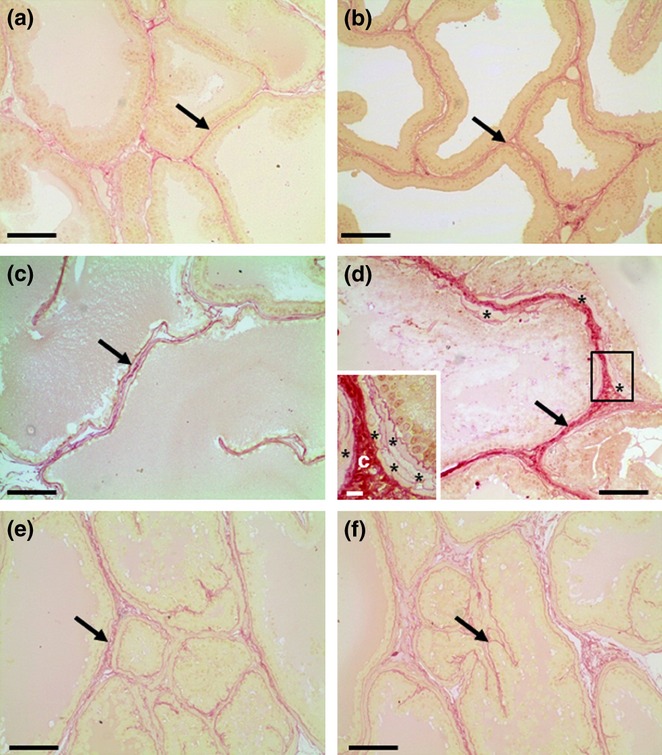
Picrosirius-stained sections of control (a, e) and caffeine-treated (b, c, d) prostatic lobes. Ventral (a–d) and dorsal (e–f) prostates. Collagen fibers (arrows) are observed around acini, mainly in the interstitial space. (b) Caffeine-treated prostate presenting area with similar morphology and collagen distribution (arrows) than control group. (c) Caffeine-treated prostate showing enlarged acini and regular collagen fibers (arrow) distribution in the reduced stroma. (d) Hyperplasic area of caffeine-treated prostate showing increased amount of collagen fibers (arrow) in the interstitial space and thickened layer of smooth muscle cells (asterisks). Insert: Detail of the figure d (square) showing thickened collagen fibers (c) and smooth muscle cells layer (asterisks). Scale bars: a–f = 50 μm; Insert = 10 μm.