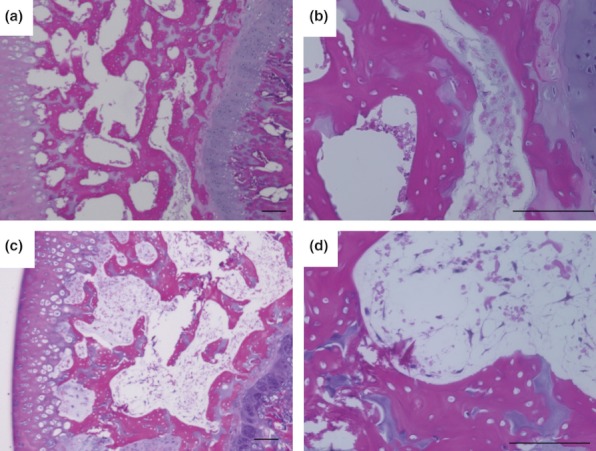
Figure 2.

Histological appearance of femoral head osteonecrosis. Haematoxylin and eosin staining of the femur. Typical specimens of weight-bearing (WB) (a, b) and non-weight-bearing (NWB) (c, d) rats. Both WB and NWB group shows diffuse presence of empty lacunae in the bone trabeculae, accompanied by surrounding bone marrow cell necrosis at most areas of femoral head. Scale bar = 100μm.
