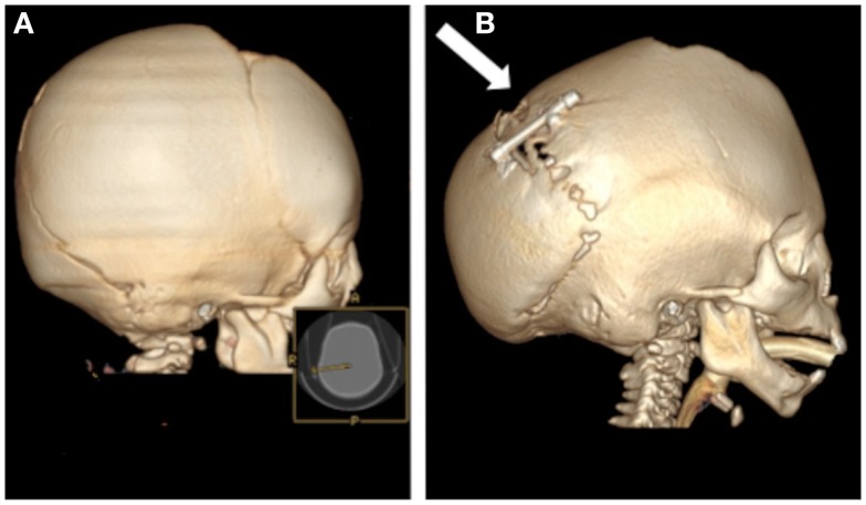
Figure 3.
Computed tomography scans of an infant with multiple suture synostosis preoperatively (A) and 4 months after distraction of the posterior cranial vault (B). The distraction footplates have been gradually separated by a distance of 25 mm and evidence of calcified bony regenerate is present between the footplates (arrow).

