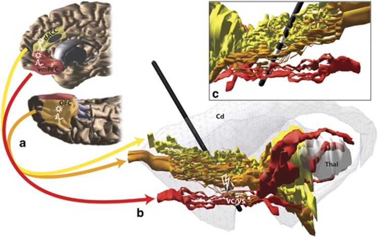
Figure 1.
Schematic illustrating key cortical areas involved in obsessive-compulsive disorder (OCD) and their pathways through the internal capsule. (a) Red, orange, and yellow fibers originate in ventromedial prefrontal cortex (vmPFC), orbitofrontal cortex (OFC), and dorsal anterior cingulate (dACC), respectively. The approximate cingulotomy site is depicted as a dark gray oval. (b) Medial view of a sagittal section with vmPFC, OFC, and dACC fibers passing through the internal capsule. The striatum is indicated in gray. A model of the electrode with its four contacts is placed at ventral internal capsule (VC)/ventral striatum (VS) deep brain stimulation (DBS) site. (c) Lateral view of a sagittal section shows the four electrode contact points in the VC/VS. VmPFC and OFC fibers pass through the ventral contacts, and OFC and dACC fibers pass through the dorsal contact points (Lehman et al, 2011). Action potential symbols indicate possible regions with changes in firing rates and/or local field potentials following HFS in the VS (McCracken and Grace, 2009); stars indicate regions with possible changes in plasticity following HFS to VS (Rodriguez-Romaguera et al, 2012).

