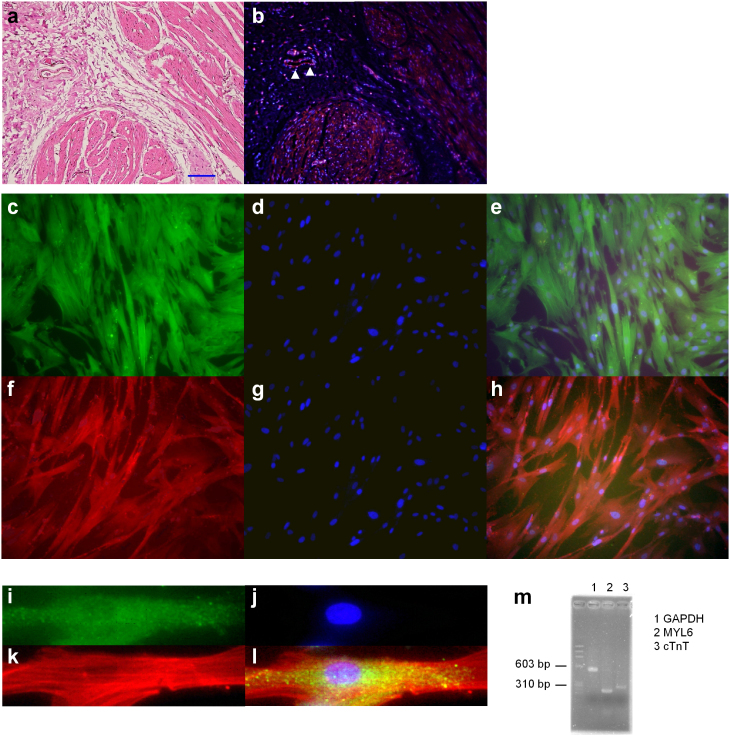
Figure 2. Histological analysis.
(a and b) A pair of thin sections of human urinary bladder detrusor smooth muscle were used. Hematoxylin-eosin (HE) staining in (a), and fluorescence of cTnT (red) and nuclear (blue) in (b). The scale bar indicates 150 μm (a), (c–l) Cultured smooth muscle cells of human detrusor smooth muscle were used in fluorescence photomicrographs. (c–e) Double staining of cTnT (green, c) and nuclear (blue, d); merge (e). (f–h) Double staining of tropomyosin (TM) (red, f) and nuclear (blue, g); merge (h). (i–l) Triple staining of cTnT (green, i), nuclear (blue, j) and tropomyosin (TM) (red, k) are shown expanded; merge (l). (m) The expression of GAPDH, MYL6 (smooth muscle marker) and cTnT in the same cultured smooth muscle cells was examined using RT-PCR.

