
Figure 12.
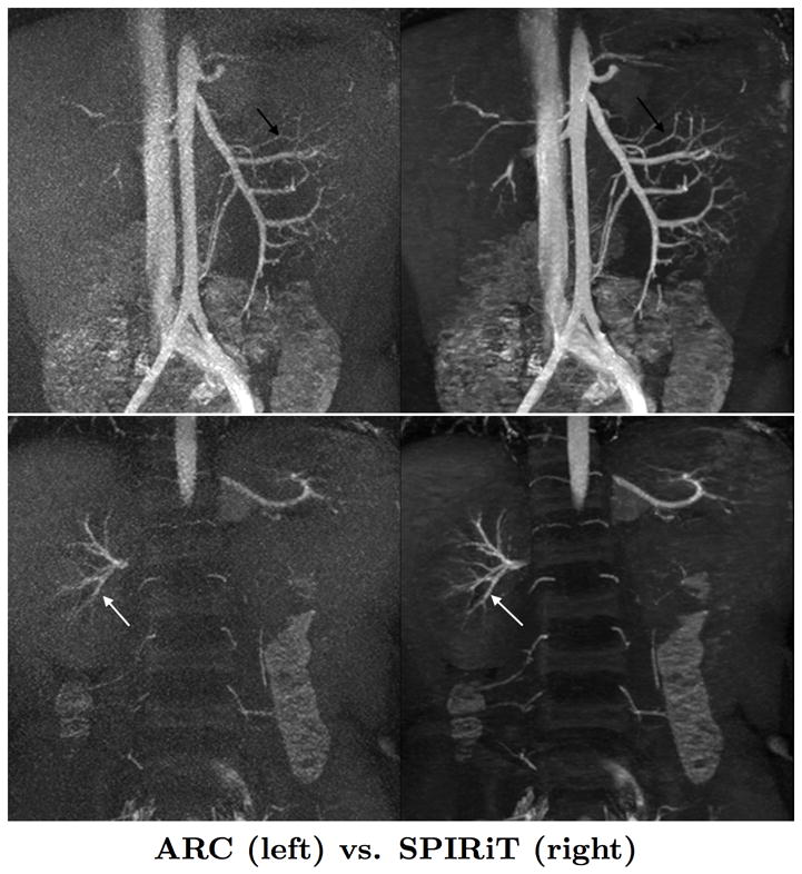
Image quality comparison of GE Product ARC (Autocalibrating Reconstruction for Cartesian imaging) [2] reconstruction (left images) with our ℓ1-SPIRiT reconstruction (right images). These MRA images of a 5 year old patient were acquired with the 32-channel pediatric torso coil, have FOV 28 cm, matrix size 320 × 320, slice thickness 0.8 mm, and were acquired with 7.2× acceleration, via undersampling 3.6× in the y-direction and 2× in the z-direction. The pulse sequence used a 15 degree flip angle and a TR of 3.9 ms. The ℓ1-SPIRiT reconstruction shows enhanced detail in the mesenteric vessels in the top images, and and the renal vessels in bottom images.