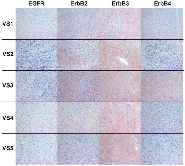
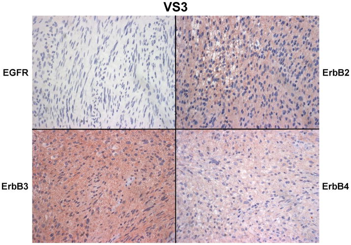
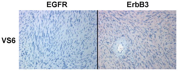
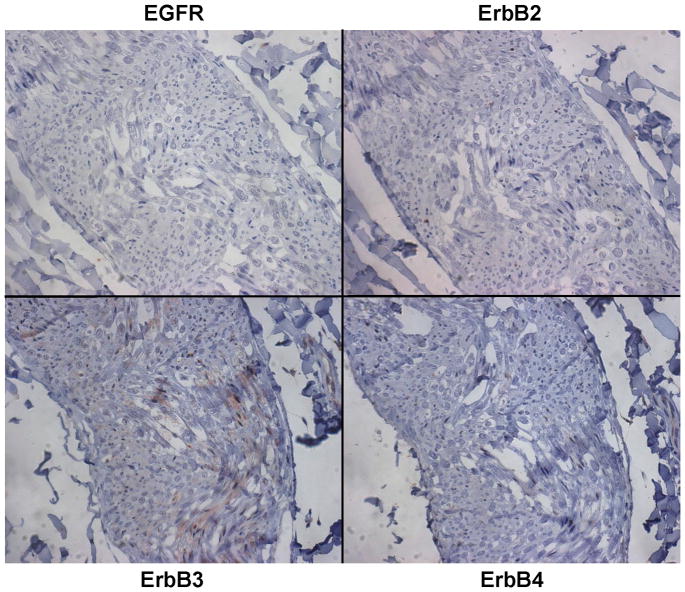
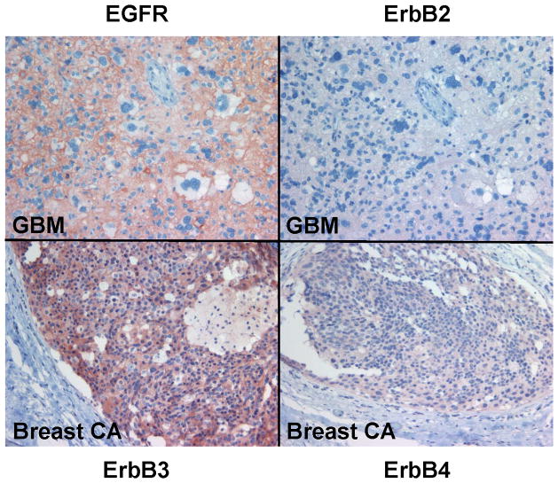
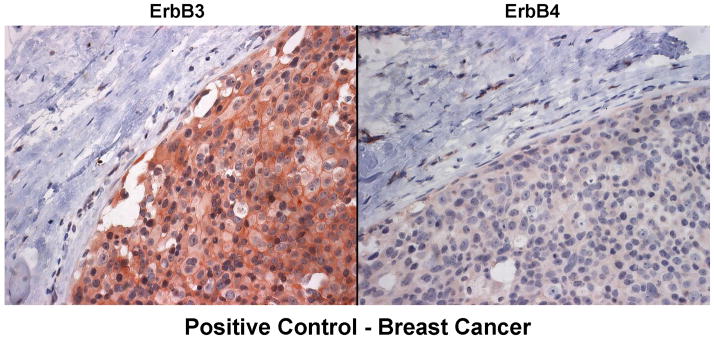
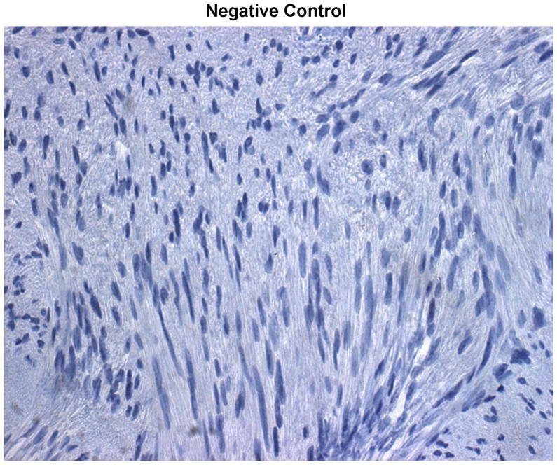
Figure 5. Immunohistochemistry analysis of VS tumor sections for ErbB protein expression.
Sections of six VS tumors were stained for expression of various ErbB members. Representative images of five VS tumor tissues immunostained for various ErbB members were shown (A). High-magnified (400x) images of stained VS tumor #3 were illustrated (B). A sixth VS tumor was also stained for EGFR and ErbB3 (C). Sections of normal sciatic nerve were also stained for expression of various ErbB proteins (D). Glioblastoma and breast carcinoma were used as positive controls for EGFR/ErbB2 and Erb3/Erb4, respectively (E). High-magnified (400x) images of beast cancer tissues stained with ErbB3 and ErbB4 antibodies were also shown (F). A VS tumor section subjected to immunostaining procedures but without the primary antibody was used as a negative control (G).

