Figure 1.
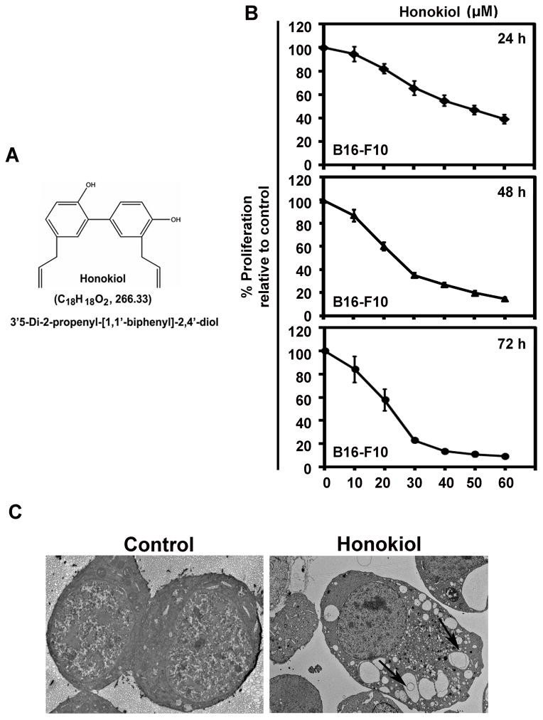
Effect of Honokiol on melanoma cancer cells. A) Chemical structure of honokiol. B) B16-F10 cells were incubated with increasing doses of honokiol (0–50 μM) at different time intervals (24, 48 and 72h) to determine the cell proliferation of B16-F10 cells. The honokiol treatment resulted in a significant dose and time-dependent decrease in cell proliferation compared to their respective controls (P < 0.05). C) Transmission electron micrograph showing honokiol induced vacuoles formation in B16-F10 cells at 24 h of honokiol (30 μM) treatment. All images were taken at 2000 magnification.

