
Figure 5. Anomalous lymphocyte localization in helminth-challenged Rgs16–/– lungs.
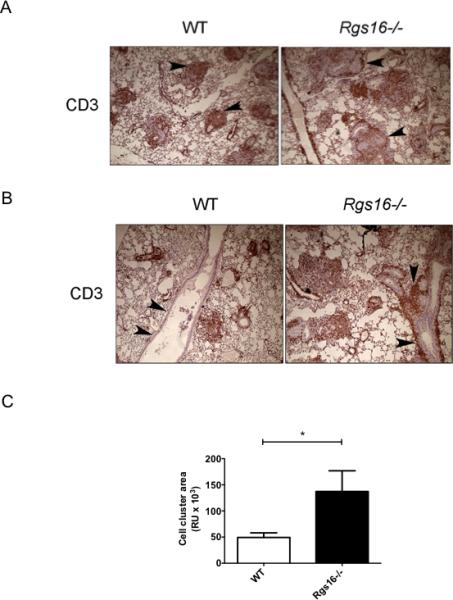
(A-B) T cell localization in helminth-challenged lungs evaluated by immunohistochemistry with a CD3 antibody. Images in (A) show parenchymal T cell accumulation in granulomas while those in (B) demonstrate perivascular/peribronchial cell aggregates (arrowheads). Total area containing cellular aggregrates around airways and vessels was quantified using Image Pro Plus software (*P = 0.04, unpaired t-test) as indicated in (C). Images represent 8 mice/group evaluated in 2 independent experiments.