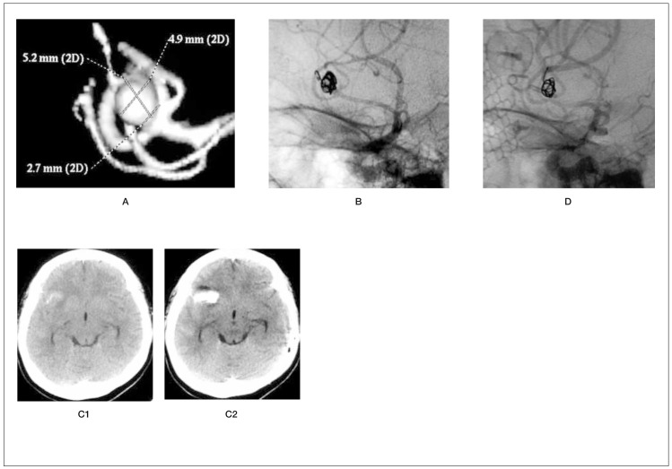
Figure 1.
A) preoperative 3D-DSA of a right middle cerebral artery aneurysm. B) Protrusion of end of first coil and microcatheter from the aneurysmal dome. C1) Preoperative CT scan. C2) CT scan immediately after perforation demonstrates intracerebral haematoma. D) DSA following clipping surgery. Thirty-seven-year-old woman, Hunt and Kosnik grade 2. The perforation of the dome of the right middle cerebral artery aneurysm occurred at the end of the first-coil insertion. Avoiding immediate removal of the coil and microcatheter, heparin reversal was performed. Following the detachment of the first coil, the insertion of the second coil from the outside into the inside of the perforated dome was attempted, but failed. After this procedure, extravasation was identified. Brain CT scan revealed an intracerebral haematoma. Subsequently, clipping surgery was performed successfully. She was discharged 31 days after the clipping surgery without any neurological deficits.

